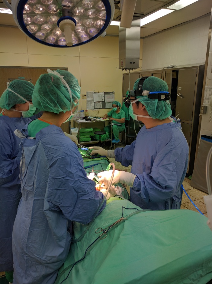
Today the operation room schedule was jam packed with orthopedic surgeries, many involving the removal of implants (ROI) from previous surgeries.
The first one I watched was the removal of pins from a patient’s patella. The patient had shattered his patella ten years ago, and was now presenting with knee pain. It was a very labor intensive procedure performed by Dr. Shih, who had to use a lot of force pulling the pins out.

I then attempted to watch a spinal fusion surgery for an elderly woman who presented with herniated bulging discs from L3-L5. Those discs would be fused anteriorly (Anterior Lumbar Interbody Fusion), which would serve to achieve posterior decompression. The surgery seemed extremely complicated as the table was raised very high and it required a great number of bodies present in the room, so I could not get a clean view of the procedure.


In the afternoon, I requested to watch a total knee replacement/arthroplasty for a 75 year old woman with osteoarthritis who had her left knee replaced just 3 months prior. This was probably the gnarliest surgery I had ever watched. What surprised me most was that the patient was awake for the entire procedure, having been anesthesized via spinal tap.

The procedure was performed by Dr Shih’s father, who was previously a director at the Taiwan Hospital Orthopedic Surgery Department, and said he had performed over 3,000 total knee replacements in his career. The surgery really reminded me of carpentry due to the true craftsmanship required by the surgeon.
The first step was to saw of the parts off the surfaces of the bone which showed osteoarthritis. The electric saw was guided by a metal guide which was precisely nailed to the distal femur to ensure proper and straight cutting. After sawing, debris was carefully cleaned off the joint space and edges of the bone.

After this step, the surgeon was grabbed a variety of tools to remove the ligaments of the knee (chiefly, the ACL, PCL, and MCL). They also had signs of wear and tear and would actually serve no purpose in an arthroplastic knee. Along with the ligaments, the lateral and medial menisci were also removed, which would be replaced by a articulatory spacer.

Dr. Shih then proceeded to saw off the proximal tibia. After sawing, there were temporary prosthetics implanted to make sure the spacing, and alignment were 100 percent accurate. There was even a special tool used to make sure the tibia prosthetic was not internally or externally rotated from the tibial tuberosity.
After everything was checked and aligned, the actual prosthetics were cemented into the bone. It was actually quite beautiful how natural the knee seemed with the implants. When these bones were fixed, the last missing part of the puzzle was the patella, which also had its internal surface sawed and placed with a prosthetic metal part. Again, I could not believe the patient was awake for the entire procedure. The sounds of the hammering and sawing would be enough to make me sick to my stomach were I in her position.
My day ended with Dr. Zhuang giving me a comprehensive lecture on the foundation of orthopedics, which mainly focused on musculoskeletal injuries and clinical tests used to confirm diagnoses. As we went through his 208 slide powerpoint, he would sometimes interject and talk about his own experiences as a student and resident, many times complaining about the long list of names given to injuries and fractures, and the rare times he would encounter certain conditions not common in Taiwan like juvenile rheumatoid arthritis. The lecture was made more enjoyable due to Dr. Zhuang’s great sense of humor.